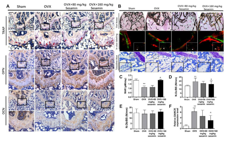
Figure 7.
Histomorphometry of the distal femur from OVX mice upon sesamin treatment. (A) Representative images of femoral metaphysis with IHC staining using TRAP, OCN and OPN antibodies. Red arrow indicates the TRAP positive cells. Scale bar: 200 μM. (B) Von Kossa staining, in vivo double labels and Toluidine blue staining of the distal femur sections. Scale bar: 400 μM for Von Kossa, 100 μM for in vivo labeling and Toluidine blue staining. (C–E) Histomorphometric analysis of distal femur sections including MAR (mineral apposition rate) (C), N.Oc/BS (number of osteoclast per bone surface) (D) and N.Ob/BS (number of osteoblasts per bone surface) (E). (F) Serum level of DANCR was revealed in OVX mice upon sesamin treatment. Data were shown as mean ± SD (n = 8; ** p < 0.01, versus sham group; ^ p < 0.05. versus OVX group).

