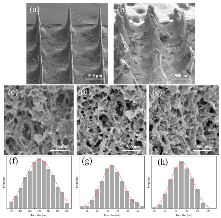
Figure 8.
SEM images of the surface and section morphology of MT/SF@MNs: (a,b) surface images before and after microneedle swelling of Pro/MT/SF = 10/1/100, (c–e) cross-section images of Pro/MT/SF = 10/1/100, 20/1/100, 30/1/100 after microneedle swelling, and (f–h) pore size frequency distribution datagrams of Pro/MT/SF = 10/1/100, 20/1/100, 30/1/100 after microneedle swelling.

