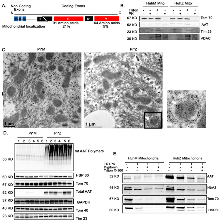
Figure 5.
AAT association with hepatocyte mitochondria. (A) Alignment of the sequences of amino acids in the AAT molecule. (B) Western blot analysis showing sequential Triton and proteinase K treatments of the mitochondrial fraction of MAAT- and ZAAT-expressing Huh7.5 cells. A voltage-dependent anion-selective channel (VDAC) was loaded as mitochondrial loading control. (C) Immuno-electron microscopy results determining the morphology and localization of the mitochondria in the liver tissue from Pi*M and Pi*Z transgenic mice. Immunogold-labeled antibody against hAAT was used to detect mitochondrial hAAT (white arrow). ER entrapped between lipid droplets and mitochondria is indicated by the black arrow. (D) Native, non-denaturing Western blot analysis of hepatic mitochondrial ZAAT aggregates in the 6 Pi*M (1–6) and 6 Pi*Z (1–6) mouse liver tissue as well as the total levels of AAT, mitochondrial markers, and GAPDH as loading control. (E) Western blot analysis of isolated mitochondrial fraction of Huh7.5 cells, showing the exact localization of AAT in permeabilized mitochondria using proteinase K, digitonin, and Triton X-100.

