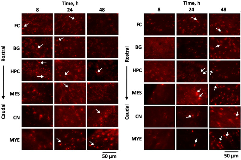
Figure 6.
Localization of the negatively charged NPs (left panel) and positively charged NPs (right panel) after IN administration. Frontal cortex (FC); basal ganglia (BG); hippocampus (HPC); mesencephalon (MES); cerebellar nuclei (CN); myelencephalon (MYE). The white arrows indicate the presence of rhodamine-labeled NPs. Adapted with permission from [144], Elsevier, 2017.

