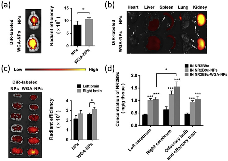
Figure 10.
Biodistribution of WGA-NPs and NR2B9c loaded into WGA-NPs IN in the rats with stroke model. (a) Biodistribution of DiR-labeled NPs and WGA-NPs in brain. Ex vivo fluorescence brain images (left) and bar graft (right, n = 4). (b) Ex vivo fluorescence imaging of peripheral organs. (c) Fluorescent images of brain slices (left) and bar graph (right, n = 4) (d) Biodistribution of NR2B9c at 1 h after intranasal administration with NR2B9c, NR2B9c NP or NR2B9c-WGA NP in different brain areas of rats. A, C and D: * p < 0.05, between groups; D: *** p < 0.001, when compared with NR2B9c IN in the same brain area. Reproduced with permission from [226], Elsevier, 2019.

