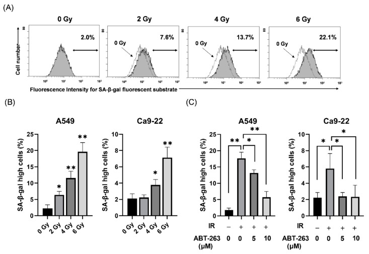
Figure 1.
Effect of ABT-263 on populations of SA-β-gal high-activity cells in irradiated cancer cells. (A) X-irradiated A549 cells were cultured for 48 h and SA-β-gal activity was analyzed using the Senescence β-Galactosidase Activity Assay Kit. Representative histograms of SA-β-gal activity in A549 cells are shown. The dotted line shows the results of non-irradiated cells and the inset numbers show the percentage of SA-β-gal high-activity cells. (B) X-irradiated A549 and Ca9-22 cells were cultured for 48 h prior to analysis of SA-β-gal activity. The results are shown as the percentage of SA-β-gal high-activity cells. (C) A549 and Ca9-22 cells irradiated at 6 Gy were cultured for 24 h prior to the addition of ABT-263 to the culture. After culturing (48 h for A549 and 24 h for Ca9-22), SA-β-gal activity was analyzed. The results are shown as the percentage of cell with high SA-β-gal activity. Data are presented as the mean ± standard deviation (SD) of independent experiments. * p < 0.05, ** p < 0.01.

