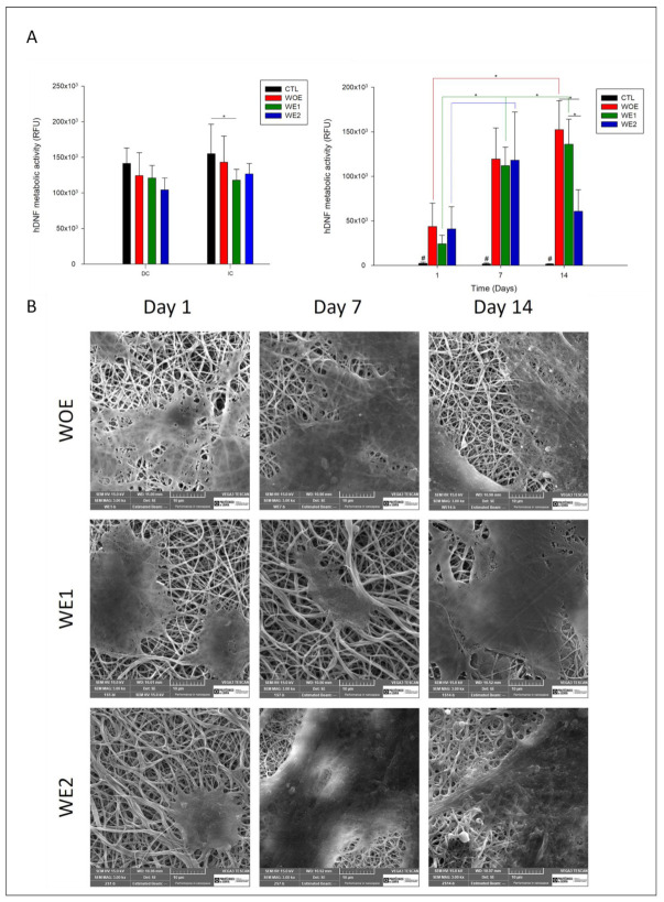
Figure 5.
(A) On left: Cytotoxicity of electrospun meshes containing phlorotannins-enriched extract (WE1-green bar; WE2-blue bar) and without extract (WOE-red bar) were evaluated by direct (DC) and indirect contact (ID) with a positive control (black bar) of viable cells. Data are expressed as the mean ± SD. Multiple group comparisons were performed using t-test with p ≤ 0.05 (*), n = 4. On right: Proliferation of hDNF cells on WOE meshes (red bars), on WE1 meshes (green bars), and WE2 (blue bars), assessed for 1, 7, and 14 days compared with the control meshes (without cells seeded; negative control–black bar). For each time point and multiple groups ANOVA one-way p ≤ 0.05 (*) was used, “#” for statistical significance compared with all other samples. (B) SEM micrographs representative of hDNF proliferation in different electrospun meshes over 14 days with a magnification of 3000 × (scale bar: 10 µm).

