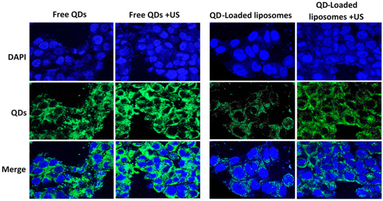
Figure 6.
HCT116 cells incubated with free QDs or QD-liposomes with and without sonication with ultrasound (US) for 1 min at 37 °C. The first row only shows the nuclei of the cells stained with DAPI (blue). An argon laser (520/50 nm) was used for QD excitation producing a green fluorescence (second row). The third row shows merged images of both the nuclei and QDs present inside the cells.

