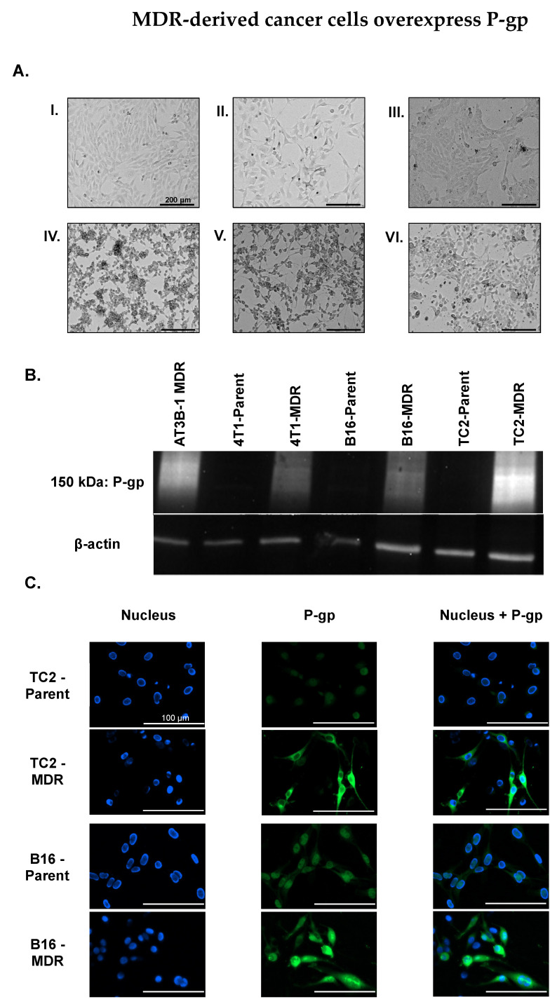
Figure 3.
(A) MDR cancer cells were derived from parent cancer cell lines by incrementally increasing the Dox concentration in growth media. (I) TRAMP-C2 (TC2) prostate parent cancer cell line. (II) B16-F10 (B16) melanoma parent cancer cell line. (III) 4T1-Luc2 (4T1) breast parent cancer cell line. (IV) TC2-MDR cancer cell line. (V) B16-MDR cancer cell line. (VI) 4T1-MDR cancer cell line. Scale: 200 µm (B) Western blot shows the increased expression of P-gp in MDR cancer cells compared to parent cancer cell lines. (C) TC2-Parent, TC2-MDR, B16-Parent, and B16-MDR stained with Anti-P-gp-Alexa Fluor 488 antibody conjugate reveals the increased expression of P-gp in MDR-derived cancer cell lines, as well as membrane localization of P-gp.

