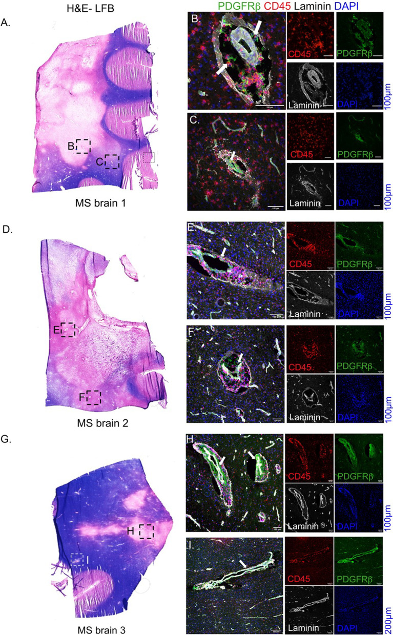
Fig. 6.
Pericytes in MS brain. Fresh-frozen MS brains (20 µm thick) harboring active lesions were investigated for pericyte morphologies. A H&E-luxol fast blue (H&E-LFB) staining of an MS brain section from a 60-year-old female showing demyelinating lesions. B, C MS brain section was stained for PDGFRβ and CD45 to locate the pericytes and infiltrating leukocytes in two different lesions demarcated as B and C in panel A. D–F MS tissue from a 61 year old male showing perivascular cuffs (CD45+ cells within laminin+ blood vessel boundaries) and intact PDGFRβ+ cells along the inflamed vasculature in two different brain regions, E and F. G Brain section from a 26 year old MS male also exhibits largely intact PDGFRβ+ staining encased within laminin+ basement membranes (H and I). Arrows show perivascular location of PDGFRβ+ pericytes in 2 regions with CD45+ leukocytes. Scale 100 µm

