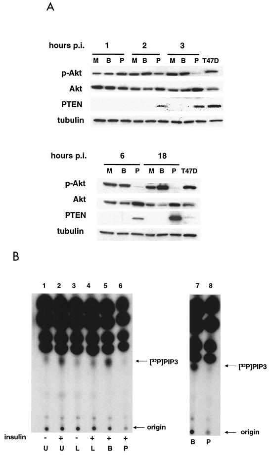
FIG. 2.
PTEN expression blocks insulin-induced production of PtdIns-3,4,5-P3 and inhibits Akt activity. (A) MDA-MB-468 cells were either mock (M), Ad-β-gal (B), or Ad-PTEN (P) infected, and total cell lysates were collected at various time points postinfection (hours p.i.) as described in Materials and Methods. T47D, total cell lysate of a breast cancer cell line expressing endogenous PTEN. Cell lysates were subjected to immunoblot analysis with phospho-Akt, Akt, PTEN, and tubulin antibodies. Tubulin antibody was used as a loading control. (B) MDA-MB-468 cells were either left uninfected or infected with Ad-β-gal or Ad-PTEN overnight. Cells were serum starved for 4 h in phosphate-free medium containing 32Pi (100 μCi/ml) before 30 min of treatment with 20 μM LY294002. Insulin (100 μ/ml) or FBS (10%) was added as indicated for 10 min. Lipids were subsequently extracted and separated on a thin-layer chromatography plate as described in Materials and Methods and visualized by autoradiography. U, untreated/uninfected; L, treated with 20 μM LY294002; B, Ad-β-gal infected; P, Ad-PTEN infected; [32P], [32P]PtdIns-3,4,5-P3.

