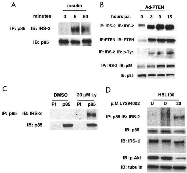
FIG. 6.
Inhibition of the PI3K pathway leads to increased association of IRS-2 and p85. (A) MDA-MB-468 cells were serum starved for 24 h and pulsed with 100 ng of insulin/ml for the indicated time periods. Cell lysates were collected and immunoprecipitations with p85 antibody were performed as described previously. The resulting immmunocomplexes were subjected to immunoblot analysis with IRS-2 and p85 antibody. IP, immunoprecipitation; IB, immunoblot. (B) MDA-MB-468 cells were infected with Ad-PTEN, and cell lysates were collected at various time points postinfection (hours p.i.). Cell lysates were immunoprecipitated with IRS-2, and the resulting immunocomplexes were subjected to immunoblot analysis with antibodies to IRS-2, p85, and p-Tyr. Also, cell lysates were immunoprecipitated with PTEN and immunoblot analysis was performed on the immunocomplexes using PTEN antibody to determine the level of PTEN expression. Total cell lysates were subjected to immunoblot analysis with p85 antibody as a control. IP, immunoprecipitation; IB, immunoblot. (C) MDA-MB-468 cells growing in medium with 10% FBS were treated with DMSO or 20 μM LY294002. Cell lysates were collected after 3 h of treatment and immunoprecipitated with p85 antibody. Immunocomplexes were subjected to immunoblot analysis with IRS-2 and p85 antibody (control). PI, preimmune serum; IP, immunoprecipitation; IB, immunoblot. (D) HBL100 cells growing in medium with 10% bovine serum were left untreated or treated with DMSO or 20 μM LY294002. Cell lysates were collected after 3 h of treatment. A portion of the cell lysates was immunoprecipitated with p85 antibody. Immunocomplexes were subjected to immunoblot analysis with IRS-2 and p85 antibody (control). Total cell lysates were subjected to immunoblot analysis with IRS-2, phospho-Akt, and tubulin (loading control) to analyze protein levels. U, untreated; D, DMSO.

