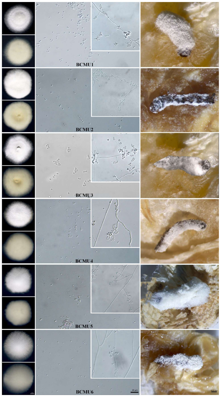
Figure 1.
Pictorial presentation of BCMU1–BCMU6 colony on the obverse and reverse sides on PDA media, conidia, and the hyphae and the mycosis caused on Spodoptera frugiperda. Scale Bar = 1 µm, 20 µm, and 0.02 mm respectively. The isolates were cultured on potato dextrose agar for 14 days at 25 ± 1 °C with a photoperiod of 12:12 h (Dark: Light). Once the larvae had died, they were placed on moist conditions to allow mycosis.

