Figure 6.

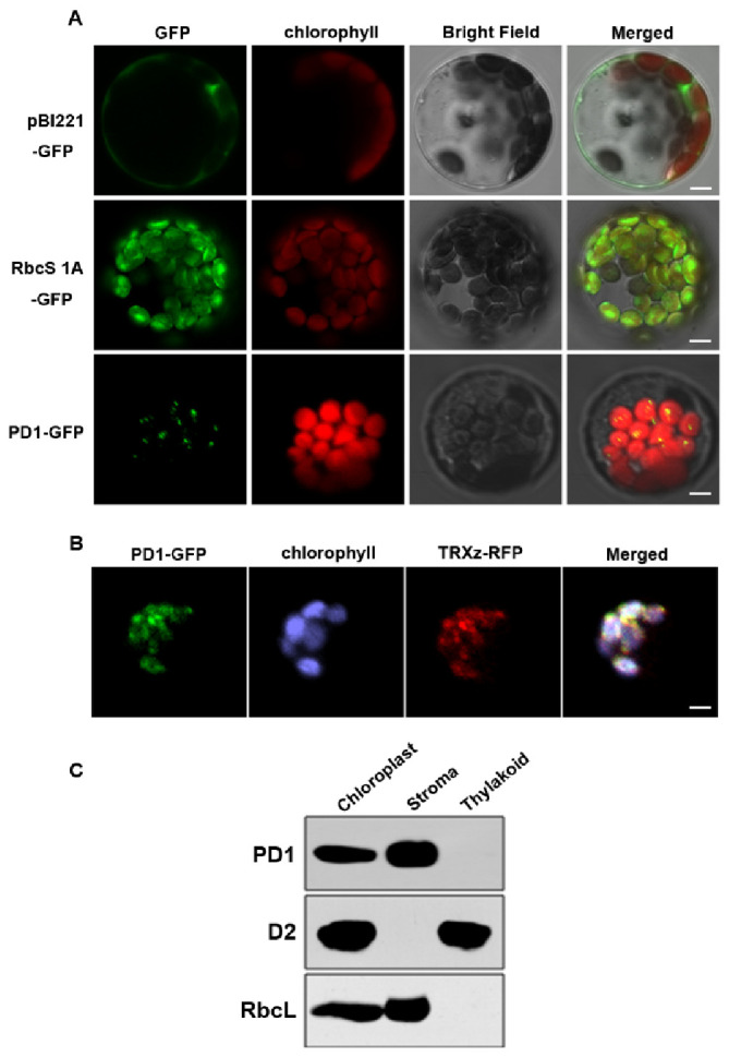
Subcellular localization of the PD1 protein. (A) Localization of PD1 protein within chloroplast by GFP assay. Chimeric proteins were transiently expressed in Arabidopsis protoplasts. pBI221-GFP, control with the pBI221 empty vector; RbcS 1A-GFP, chloroplast control; PD1-GFP, PD1-GFP fusion. Chlorophyll autofluorescence of chloroplasts is shown in red. Bars = 3 μm. (B) Colocalization of PD1-GFP with TRXz-RFP. The fluorescence signal of PD1-GFP overlaps with that of TRXz-RFP within chloroplast nucleoids. Chlorophyll autofluorescence of chloroplasts is shown in purple. Bars = 3 μm. (C) PD1 localizes in the chloroplast stroma. Intact chloroplasts were isolated from the leaves of wild type seedlings and then separated into thylakoid membrane and stroma fractions. Polyclonal antisera against the PD1, D2, and RbcL were used in immunoblot analysis.
