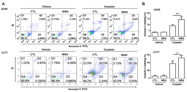
Figure 2.
WSG enhances cisplatin-induced apoptotic responses. (A) After 48 h exposure to WSG (120 μg/mL) and/or cisplatin (10 μM), the cells were subjected to co-staining with the annexin V–FITC/PI kit. Flow cytometry was performed for apoptosis analysis. The percentages of apoptotic cells in early and late apoptosis were determined using FlowJo software. (B) The data, representative of three separate experiments, are presented as means ± standard deviations; error bars reflect standard deviations. Significant differences between the treatment and control groups are presented (** p < 0.01).

