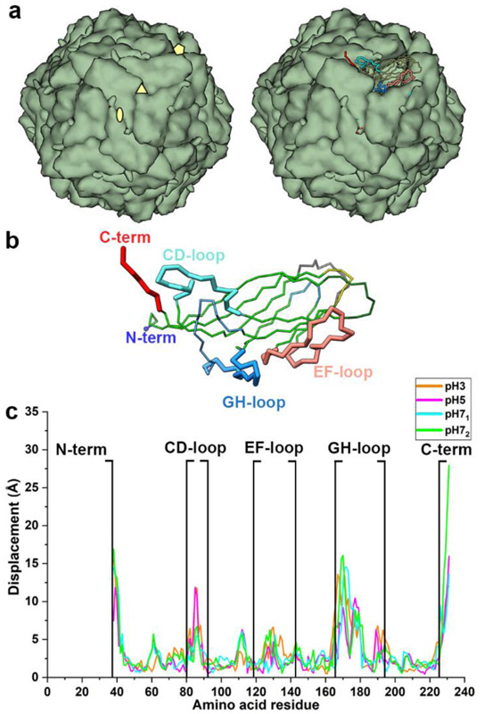
Fig. 3.
Conformational variability of CP at pH 3, 5 and 7. (a) left: PCV2 capsid with CP shown as green blobs. The icosahedral axes of symmetry (iaos) are shown as pentagon (5-fold), triangle (3-fold), and ellipse (2-fold). Right: one of the blobs (subunits) is shown as a Cα tube representation with the important loops (N-term, CD, and GH as thick tubes). (b) PCV2 CP Cα tube representation with important loops (thick tubes) labelled. (c) distances between equivalent Cα atoms of the representative conformations at pH 3, 5, 7 after alignment to capsid structure (PDB entry 3R0R). Panels a and b were made using the structure of the capsid. In the context of the assembled capsid, the N-terminus is in the interior of the capsid near the 2-fold iaos, CD-loop is located on the surface of the capsid near the 2-fold iaos, GH-loop is buried near the 3-fold iaos, and the C-terminus is located on the capsid surface near the 2-fold iaos.

