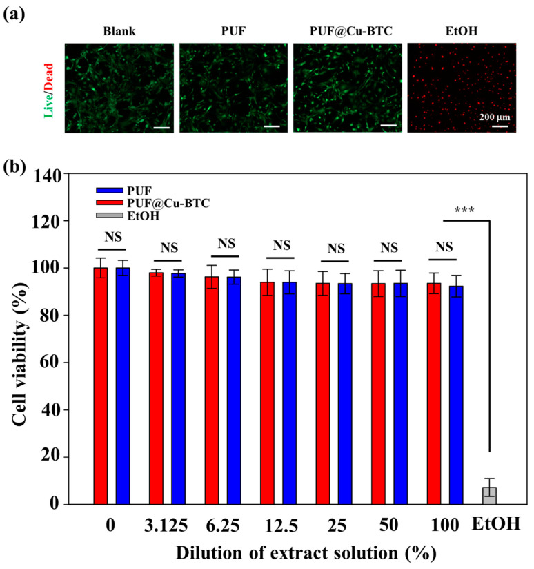
Figure 8.
(a) Live/dead staining images of Mouse Embryonic Fibroblasts (MEFs) after contact with PUF only or PUF@Cu-BTC on day 1. Positive control (blank): cells cultured with no contact. Negative control: cells contacted with EtOH. (b) In vitro cytotoxicity of extract solution of PUF only or PUF@Cu-BTC toward MEFs. (Mean ± standard deviation with n = 4; NS: not significant, *** p < 0.001. Scale bar = 200 μm).

