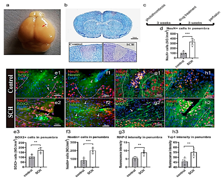
Figure 2.
SCH promoting neural regeneration in stroke model by photothrombosis. (a) Stroke model by photothrombosis. (b) Nissl’s staining of brain ischemic area (grey) at 48 h. (c) Schedule of experimental manipulations, scale bar: 1 mm. (d) Quantification of the number of NeuN+ neurons in the ischemic area. SOX2 (green), nestin (green), NeuN (red), DAPI (blue), scale bar: 50 μm; (e1) The hscattered SOX2 positive cells in the ischemic and core areas of the control group, (e2) SOX2 positive NPCs in SCH; (f1,f2) The number of nestin-positive cells; (e3,f3) The number of Nestin and SOX2 positive progenitor cells in SCH group was higher than that in the control group. (g1,g2,h1,h2) Nerve fiber regeneration in the ischemic area. MAP-2 (green), Tuj-1 (green), NeuN (red), DAPI (blue); (g3,h3) MAP-2 and Tuj-1 nerve fibers axon or dendrites grow into penumbra in SCH group. The nerve fibers density in SCH group was higher than that in the control group. Data are expressed as the mean ± SEM, and were analyzed by t test N = 5, ** p < 0.01, *** p < 0.001.

