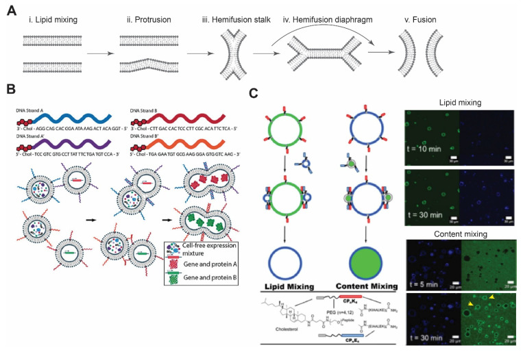
Figure 3.
Membrane fusion. (A) Schematic of lipid membrane fusion showing sequential stages from protrusion to hemi-fusion and fusion pore. Redrawn from Reference [115] with permission from Springer Nature, copyright 2008. (B) Complementary DNA strands on two different vesicles eventually lead to their fusion and allow mixing of the contents. This can be utilized to initiate any biochemical reactions, such as in vitro protein synthesis. Reproduced from Reference [121] with permission from John Wiley and Sons, copyright 2019. (C) Schematic diagram of coiled-coil peptide-mediated vesicle fusion. Peptide K and peptide E were incorporated on the surface of vesicles using cholesterol with PEG as a linker. GUVs and LUVs were used in lipid and content mixing. Appearance of florescence signals after 30 min of incubation confirmed lipid mixing, while in content mixing, release of an encapsulated dye in the lumen of GUVs was observed (yellow arrows). Reproduced from Reference [122] with permission from Springer Nature, copyright 2020.

