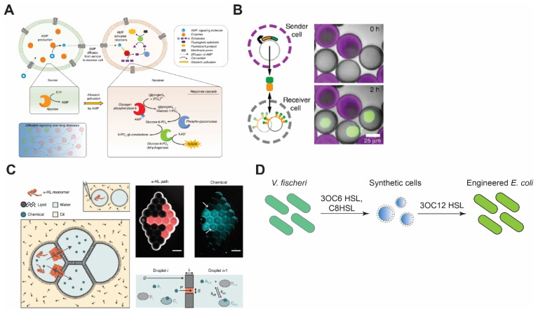
Figure 4.
Synthetic cell communication with synthetic or living cells. (A) Signal amplification via allosteric activation of glycogen phosphorylase b by AMP. Sender cells generate AMP and send it through α-HL pores. Reproduced from Reference [135] with permission from Springer Nature, copyright 2020. (B) Porous synthetic cells that contain DNA-bound clay synthesize signal molecules and send them via chemical diffusion. Receiver cells encapsulate DNA sequences that encode binding sites for the signal molecules. Reproduced from Reference [136] with permission from Springer Nature, copyright 2018. (C) Reconstitution of synthetic cell communication in a network of droplet interface bilayers. The signal propagates through α-HL (left) or diffuses across the membranes (right) and activates cell-free expression of reporter proteins. Reproduced from Reference [137] with permission from Springer Nature, copyright 2019. (D) Synthetic cell–living cell communication via homoserine lactone molecules. Synthetic cells sense the presence of V. fischeri and send signal molecules to E. coli, thereby making E. coli sensitive to V. fischeri quorum-sensing molecules. Redrawn from Reference [138] with permission from American Chemical Society, copyright 2017.

