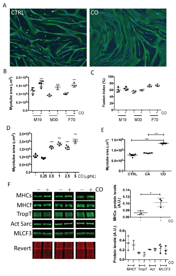
Figure 3.
Carnosol induces myotube hypertrophy. At confluence, myoblasts from different donors [M19 in (A–E) and M19, M30 and F70 in (B,C)] were induced to differentiate for three days, and then, carnosol was added (CO) or not (CTRL) to the culture at a concentration of 1 μg/mL (A–C,E,F), or at increasing concentrations, from 0.25 to 5 μg/mL (D). Two days later, myotubes were processed for (A) indirect immunofluorescence analysis with an antibody against troponin T (green). Nuclei were visualized by DAPI staining (blue). Representative images of M19 cells. (B,D,E) Quantification of troponin T images (myotube area in μm2). (C) Quantification of the fusion index (percentage). (F) Left: After cell incubation (+) or not (−) with 1 μg/mL carnosol, myotube protein extracts were analyzed by Western blotting with anti-slow myosin heavy chain (MHCs), -fast myosin heavy chain (MHCf), -troponin T (TropT), -sarcomeric actin (Act Sarc), and -myosin light chain F3 (MLCF3) antibodies. Revert® total protein stain was used as internal loading control. Right: quantification of the Western blotting data. A.U., arbitrary units (i.e., the ratio of the level of the protein of interest to the Revert® stain level. * p < 0.05, *** p < 0.001 (***). Data are the mean ± SD of three to five replicates. M19, 19-year-old male donor; M30, 30-year-old male donor; F70, 70-year-old female donor.

