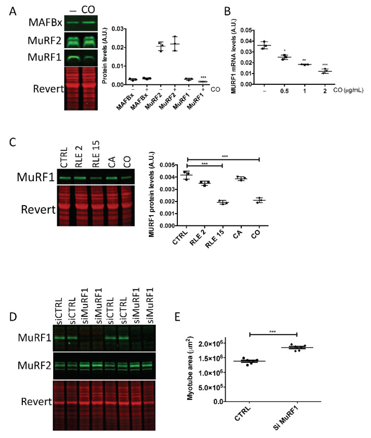
Figure 5.
Carnosol inhibits MuRF1 expression. At confluence, myoblasts (M19) were induced to differentiate for 3 days, then, (A) carnosol (CO, 1 μg/mL) or (C) rosemary leaf extract (RLE, 2 μg/mL or 15 μg/mL), carnosic acid (CA, 1 μg/mL) were added or not (CTRL) to the cultures for 48 h. Myotube protein extracts were then analyzed by Western blotting with (A) anti-MAFbx, -MuRF2 and -MuRF1 antibodies and (C) anti-MuRF1 antibodies. Revert® total protein stain was used as internal loading control. On the right, quantification of the Western blotting data; A.U., arbitrary units (i.e., the ratio of MAFbx, MuRF2 or MuRF1protein levels to the Revert® stain level). (B) RT-qPCR analysis of MuRF1 gene expression in human myotubes incubated or not (CTRL) with 0.5, 1 or 2 μg/mL of carnosol (CO) for 24 h. (D,E) Myotubes transfected with siMuRF1 and analyzed by (D) Western blotting with anti-MuRF1 and -MuRF2 antibodies, and (E) by immunofluorescence with an anti-troponin T antibody to quantify the myotube area (in μm2); * p < 0.05, ** p < 0.01, *** p < 0.001. Data are the mean ± SD of three to seven replicates. M19, 19-year-old male donor.

