Figure 1.
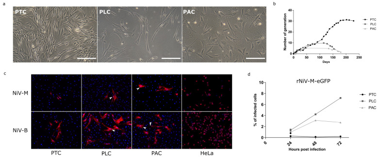
In vitro culture and Nipah viruses infections of bat primary cells (bPCs). (a) bPCs derived from the trachea (PTC), lung (PLC), and alary membrane (PAC) were observed under a light microscope. (b) Comparative growth curves of bPCs. (c) bPCs and HeLa cell line, used as control of infection, were infected with NiV-M and NiV-B isolates at an MOI of 3. At 24 h post-infection, the infected cells were visualized by NiV nucleoprotein immunostaining (red). DNA was counterstained with DAPI (blue). (d) bPCs were infected with the recombinant NiV-M virus expressing eGFP protein: rNiV-M-eGFP at an MOI of 3. The percentage of infected cells was quantified at 24, 48, and 72 h post-infection measuring GFP by flow cytometry.

