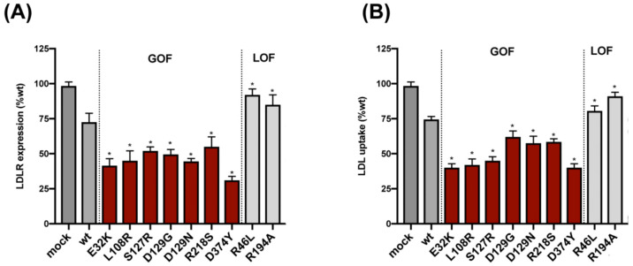
Figure 2.
Extracellular activity of PCSK9 variants on HepG2 cells. (A) Cell surface LDLR expression was determined in HepG2 cells incubated with the purified variants by flow cytometry. (B) LDL cellular uptake was determined in HepG2 cells incubated with the purified variants by flow cytometry. Histograms represent the mean ± SD of three independent experiments performed by triplicate, * p < 0.01 compared to wild-type PCSK9.

