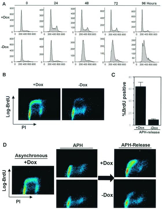
FIG. 2.
Active RB inhibits S-phase progression. (A) A2-4 cells were synchronized in quiescence by culture in medium containing Dox and 0.1% FBS for 72 h. In half of the cultures PSM-RB expression was induced by changing to 0.1% FBS medium lacking Dox for 24 h. Cells were then serum stimulated (10% FBS) in the presence or absence of Dox and harvested at the indicated time points. Cells were fixed and stained with propidium iodide and analyzed by flow cytometry. Shown are representative histograms of 10,000 gated events from two independent experiments. (B) Asynchronously growing A2-4 cells were cultured in the presence or absence of Dox for 16 h. Cells were pulse-labeled with BrdU for 1 h and then processed for bivariate flow cytometry. Two-dimensional contour maps are shown, with BrdU incorporation on the y axis and DNA content (propidium iodide [PI]) on the x axis. Data shown are representative of two independent experiments. (C) A2-4 cells were synchronized in early S phase with APH. PSM-RB expression was then induced by culture in the absence of Dox (see Materials and Methods). Cells were released from APH (see Materials and Methods) and labeled with BrdU for 4 h. Cells were fixed and immunostained for BrdU incorporation to monitor S-phase progression. Data shown are from two independent experiments with at least 150 cells counted per experiment. (D) A2-4 cells were synchronized in S phase with APH and then cultured for an additional 16 h in APH with or without Dox. Cells were then released from the APH block and pulse labeled with BrdU for 1 h. Cells were harvested and processed for bivariate flow cytometry. Shown are representative contour maps with BrdU (y axis) and propidium iodide (PI, x axis) staining.

