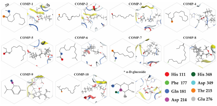
Figure 12.
Two-dimensional and three-dimensional interaction representation of the selected 10 inhibitors having a reference substrate (alpha-D-glucose) with the active pocket residues of the α-glucosidase protein. The interactive atoms of each compound with a specific residue are colored as the residue assigned color in the 2D representation. * The alpha-glucosidase protein substrate (α-D-glucose) interaction with active site residues.

