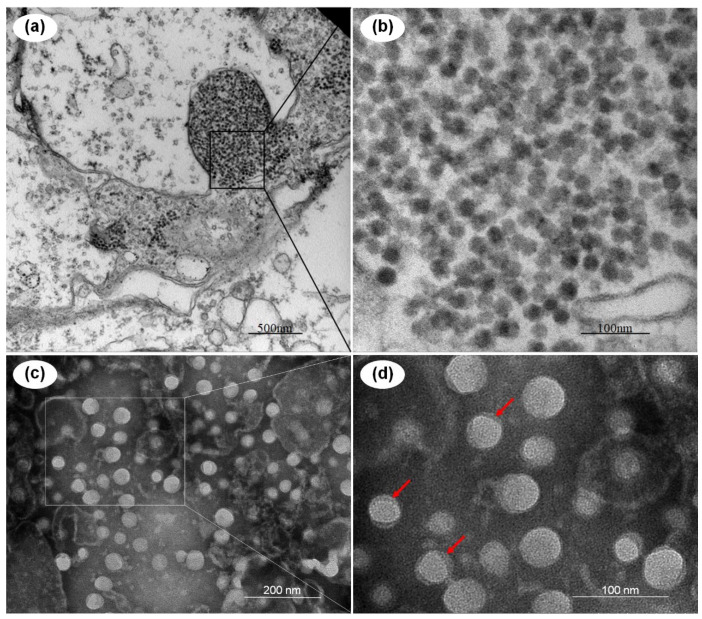
Figure 1.
Transmission electron micrographs of the Penaeus vannamei picornavirus (PvPV) virions and an ultrathin section of the hepatopancreatic epithelial cells of the moribund Penaeus vannamei from the farm in Shandong, China. (a,b) Ultrathin section of hepatopancreatic epithelial cells; (c,d) purified PvPV virions; (b–d) show magnified micrographs in the corresponding framed areas of (a–c), respectively. Note the tegument of PvPV particles, indicated with red arrows. Scale bars: (a) 500 nm, (b) 100 nm, (c) 200 nm, (d) 100 nm.

