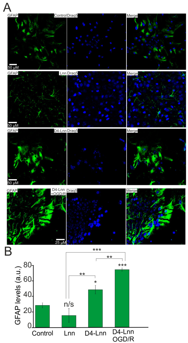
Figure 8.
Pre-incubation of cortical cells with 10 µM D4-Lnn for 24 h causes an increase in GFAP protein expression. The reactivation effect persisted 24 h after OGD/R: (A) Immunostaining cortical cells with antibodies against GFAP in the control, after 24-h pre-incubation with 10 µg/mL D4-Lnn, 10 µg/mL Lnn, and after 40 min OGD with 24-h reoxygenation (cells 24-h pre-incubated with 10 µg/mL D4-Lnn before OGD/R). Draq5—nuclei staining. (B) Intensity levels of GFAP were determined by confocal imaging. We analyzed individual cells which had fluorescence of secondary antibodies. The quantitative data reflecting the level of GFAP expression are presented as fluorescence intensity values in summary bar charts (mean +/− SEM). The values were averaged by 150 cells for each column. The results obtained after immunostaining agree well with the data of fluorescent presented in panels (A). Each value is the mean ± SE (n ≥ 3, p < 0.05). Statistical significance was assessed using one-way ANOVA followed by the Tukey–Kramer test. n/s—data not significant (p > 0.05), * p < 0.05, ** p < 0.01, *** p < 0.001. For repeats, 4 separate cell cultures were used. N (number of animals used for cell cultures preparation) = 4.

