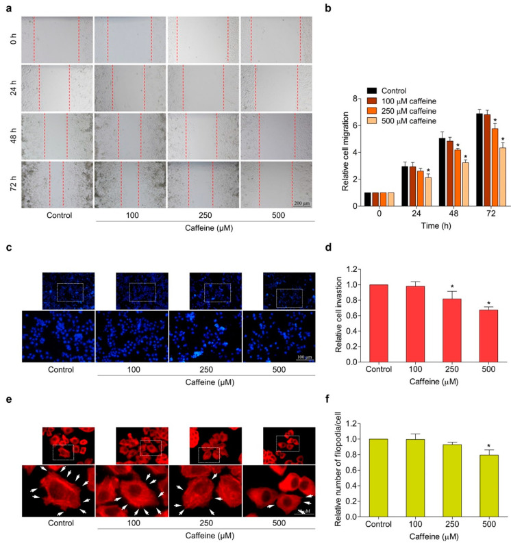
Figure 3.
Effect of caffeine on cell migration, invasion, and filopodia formation in NCI-H23 cells. (a) Cell migration activity was assessed by wound-healing assay. The cells were treated with non-toxic doses of caffeine, and the movement of cells into the wound space was evaluated at 0, 24, 48, and 72 h. (b) The cell migration rate was represented as a relative value. (c) Cell invasion was determined with the transwell Boyden chamber. After incubating with caffeine for 24 h, the invaded cells were stained with Hoechst 33342 and determined by fluorescence microscopy. (d) Relative cell invasion was calculated from the number of invaded cells in the treatment groups divided by the control group. (e) The cells were cultured with caffeine for 24 h before staining with phalloidin–rhodamine. Filopodia formation was visualized by fluorescent microscopy and is indicated by white arrows. (f) The number of filopodia per cell was calculated as a relative value. Data are presented as mean ± SD (n = 3). * p < 0.05 compared with the non-treated control.

