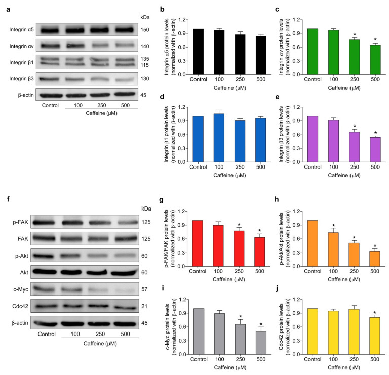
Figure 5.
Effect of caffeine on the expression of metastasis-related proteins in NCI-H23 cells. (a,f) The cells were cultured with caffeine at concentrations of 0–500 µM for 24 h. The protein expression levels of integrins α5, αv, β1, β3, phosphorylated FAK (Tyr397), total FAK, phosphorylated Akt (Ser473), total Akt, c-Myc, and Cdc42 were investigated by western blot analysis (b–e,g–j). The relative protein levels were quantified by densitometry and normalized with β-actin to confirm the equal loading of samples. Data are presented as mean ± SD of at least three independent samples. * p < 0.05 compared with the non-treated control.

