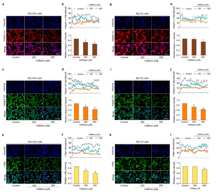
Figure 6.
Effect of caffeine on the expression of metastasis regulatory proteins in both human lung cancer cells (NCI-H23 cells) and primary human lung cancer cells (MLC15 cells). The immunofluorescence assay was performed for analysis. (a–f) NCI-H23 cells or (g–l) MLC15 cells were treated with caffeine at concentrations of 0–500 µM for 24 h, then incubated with primary antibodies (integrin αv, integrin β3, and c-Myc) followed by co-staining with Alexa Fluor 594-labeled secondary antibody (for integrin αv; red) or Alexa Fluor 488-labeled secondary antibody (for integrin β3 and c-Myc; green) and Hoechst 33342. Confocal images were visualized under fluorescence microscopy and the fluorescence intensity was measured with ImageJ software. The histogram represents the relative intensity value of the cells. Data are expressed as mean ± SD of at least three independent experiments. * p < 0.05 compared with the non-treated control.

