
FIGURE 6.
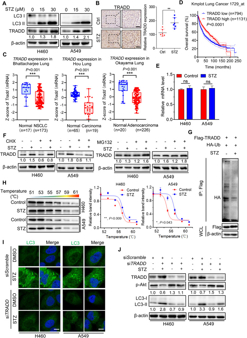
Sertaconazole induces autophagy by stabilizing TRADD in NSCLC cells. (A) Immunoblotting of TRADD expression in A549 and H460 cells treated with sertaconazole for 24 h. (B) IHC staining of TRADD in tumor xenografts. Scale bar, 50 μm. (C) Data represent the Z‐score of TRADD from Oncomine database. (D) Kaplan–Meier analysis of NSCLC patients from Kmplot database. (E) The relative mRNA levels of TRADD in cells treated with or without sertaconazole for 24 h was determined by qPCR analysis. (F) Immunoblotting analysis of TRADD protein level in cells treated with CHX or MG‐132 in the presence or absence of sertaconazole for 24 h. (G) 293T cells were transfected with Flag‐TRADD and HA‐tagged ubiquitin plasmids and then treated with sertaconazole for 24 h. Immunoprecipitation was used to detect the ubiquitination of TRADD. (H) Cellular thermal shift assay showing target engagement of TRADD by sertaconazole in A549 and H460 cells. (I, J) H460 and A549 cells were transfected with siScramble or siTRADD, followed by treatment with sertaconazole for 24 h. Immunofluorescent analysis of endogenous LC3 puncta (I) and immunoblot analysis of LC3 turnover (J) and were performed. Scale bar, 10 μm. All experiments were repeated at least three times. Statistic method: 2‐tailed Student's t‐test. Data are means with SD. ns, not significant, *p < 0.05, **p < 0.01, ***p < 0.001