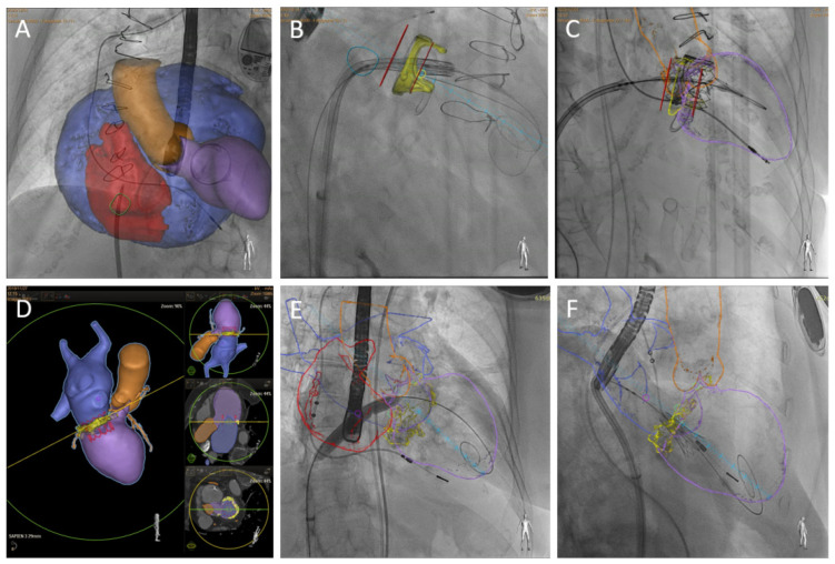
Figure 2.
CT-fluoroscopy fusion imaging. The superior row shows a TMVR valve-in-valve procedure in a patient with extreme left atrium enlargement and modified projection required for transeptal puncture (A). Markers (red lines) may be over-imposed to fluoroscopy imaging to guide depth deployment (B,C). Inferior row, TMVR valve-in-MAC CT preprocedural planning (D), interatrial septal balloon dilatation (E) and initial phase of THV deployment with coaxial projection to mitral annulus (F).

