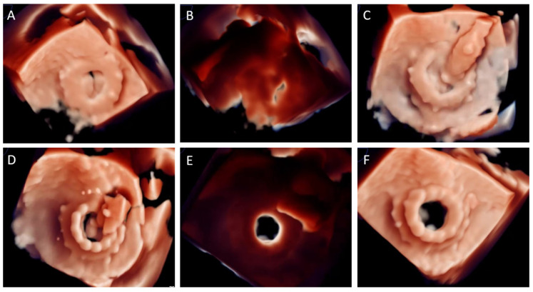
Figure 3.
Three-dimensional transoesophageal echocardiography with photo-realistic rendering during TMVR valve-in-valve procedure. (A) En-face view of a degenerated mitral surgical prosthetic valve, with severe prosthetic stenosis. (B) Same image with light source place behind mitral prosthetic valve during diastole. Prosthetic leaflets thickening and mobility reduction can be easily noted. (C) THV positioning inside SHV. (D) Balloon-expandable THV deployment. (E) Immediate result after deployment. Same image configuration than (B), significant improvement in diastolic opening can be noted. (F) TMVR ViV final result en-face view.

