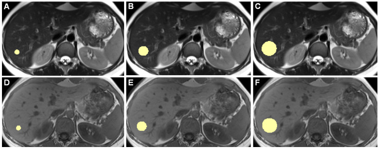
Figure 1.
Example slices of 3D ROIs of one healthy individual, acquired on the 3 Tesla I scanner, are shown. Images (A–C) are from a T2-weighted TSE HASTE sequence, (D–F) are from a T1-weighted GRE FLASH sequence. (A,D) show 10-mm diameter ROIs, (B,E) 20 mm, and (C,F) 30 mm ROIs. While drawing ROIs manually throughout all included patients, we aimed to only include hepatic parenchyma while excluding any apparent blood vessels or bile ducts.

