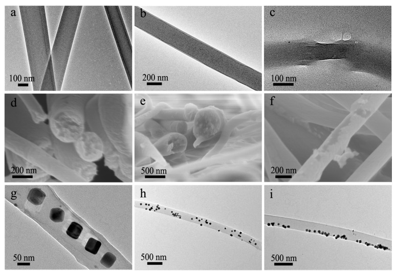
Figure 4.
(a–c) TEM images of (a,b) intact co-electrospun nanofibers and (c) a co-electrospun nanofiber with broken shell; (d–f) SEM images of the (d,e) fracture surface and (f) surface of co-electrospun nanofibers; (g–i) TEM images of Ag-NPs loaded co-electrospun nanofibers, showing the distribution of Ag-NPs in the (g) central region, (h) whole nanofibers and (i) marginal area, respectively.

