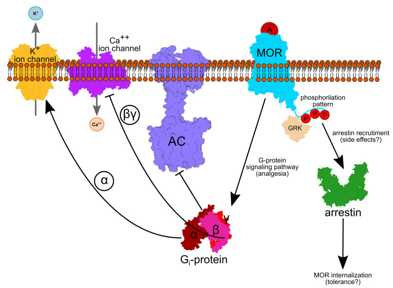
Figure 1.
The cellular path of opioid analgesia. An agonist (red circle A), such as morphine, activates the MOR. Depending on the phosphorylation (P) pattern, agonist binding leads to Gi-protein and/or arrestin-based signaling. GRK proteins add phosphate groups to specific Ser/Thr amino acid residues. An active, GTP-bound Gα inhibits Adenylate Cyclase (AC), whereas Gβγ inhibits inward Ca++ and increases outward K+ current, such that the neuronal membrane is hyperpolarized. Arrestin activation leads to internalization of the receptor, followed by degradation or recycling. Protein depictions were created with UCSF Chimera 1.14 [23], whereas the lipid membrane, text and arrows were added using Inkscape 1.0.

