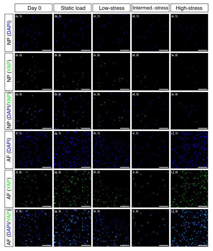
Figure 4.
Immunofluorescent pictures of the nucleus pulposus (NP) and in the annulus fibrosus (AF). The sections were stained with DAPI (blue) and anti-YAP (green) and are shown individually as well as merged. (a) NP cells at day 0, (b) NP cells after static load, (c) NP cells after low-stress load, (d) NP cells after intermediate-stress load, (e) NP cells after high-stress load, (f) AF cells at day 0, (g) AF cells after static load, (h) AF cells after low-stress load, (i) AF cells after intermediate-stress load, (j) AF cells after high-stress load. Scale bar = 100 µm.

