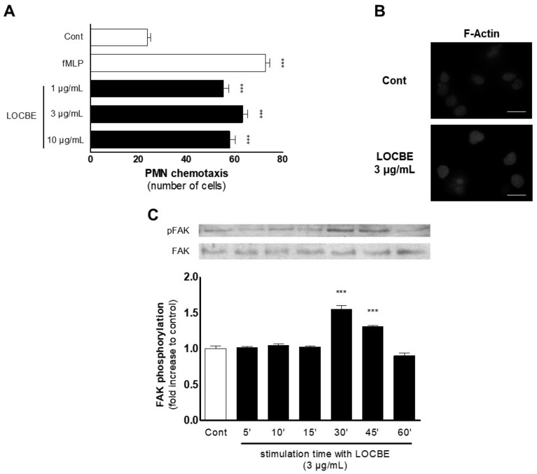
Figure 2.
LOBCE presents chemotactic activity. (A) PMNs were inserted in the upper compartment of a 48-well Boyden chamber (5 × 10⁴ cells per well) to test their migration towards LOBCE (at 1, 3, and 10 μg/mL), inserted in the lower compartment of the chamber, separated by a 5 μm pore-sized polyvinylpyrrolidone-free polycarbonate membrane. The peptide fMLP (100 nM) was used as a positive control. *** p < 0.001 compared to the control group (n = 3). (B) PMNs (106 cells per well) were incubated in the presence or absence of LOBCE (3 μg/mL) for 30 min. Then, PMNs were fixed, incubated for two hours with phalloidin-tetramethylrhodamine B isothiocyanate, and examined in a microscope equipped for epifluorescence. Scale bar: 10 μm. (C) PMNs (106 cells per well) were incubated in the presence or absence of LOBCE (3 μg/mL) for varying times (up to 60 min). Then, PMN were lysed, and extracts were run through SDS-PAGE with acrylamide gels, transferred to polyvinylidene fluoride membranes, blocked with BSA 5%, and probed with anti-pFAK or anti-actin. Immunoreactive bands were visualized using an ECL solution through a ChemiDoc Imaging System. *** p < 0.001 compared to the control group (n = 3).

