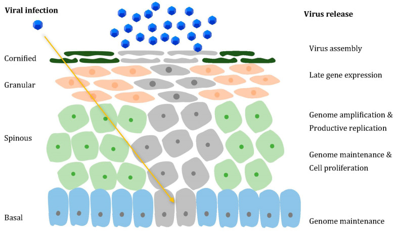
Figure 1.
HPV life cycle. Through a microscopic wound, HPV reaches the basal layer of the stratified epithelium (yellow arrow), penetrating the cells within (affected cells are pictured in grey). The infected undifferentiated basal cells ensure viral DNA replication. The productive phase is gradually activated in the suprabasal layers, consisting of increased viral genome amplification, which is ensured by the ability of the E6 and E7 proteins to promote cell cycle re-entry. In the uppermost layers, away from immune surveillance, L1 and L2 expression facilitates encapsidation, thus allowing virion assembly and release.

