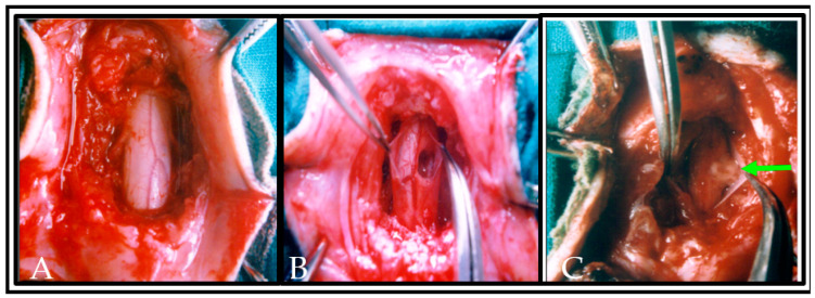
Figure 3.
Photograph showing the completion of laminectomy and exposure of the dorsal and lateral surface of normal spinal cord segments in situ with intact meningeal coverings, neighboring blood vessels and dorsal rootlets (A). In (B), the dura was slit and a hemisection lesion cavity (approximately 5 mm) was created unilaterally. Care has been taken to not disrupt the neighboring blood vessels in the left side of spinal cord or along the dorsal nerve rootlets. In (C), the hemisected lesion cavity was filled with the donor tissue and plasma clot obtained from the same animal. An arrow indicates the transplanted tissue in lesioned cavity of the spinal cord.

