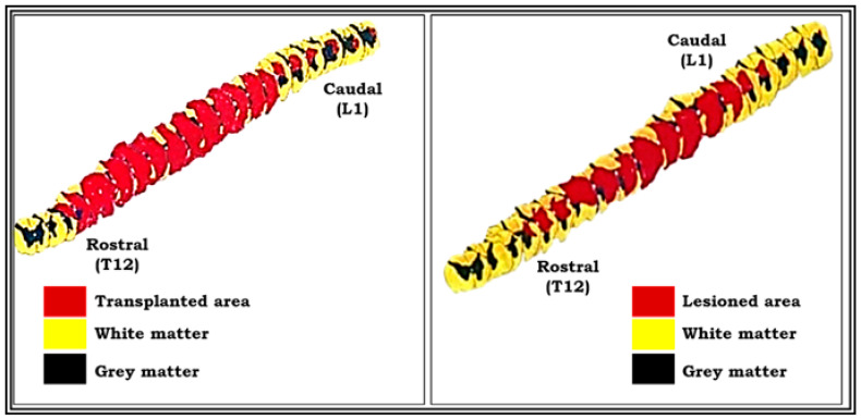
Figure 10.
Pictures showing three-dimensional graphic representation of the unilateral hemisected segment of spinal cord (pop 360 days) and transplanted segment of spinal cord (pop 360 days). Reconstruction was done on a centimeter graph paper using Nikon draw tube fitted to a Nikon labophot binocular light microscope with a magnification of 20×. This was kept constant throughout for all the serials. Red-colored part shows the lesioned area, and yellow-colored part shows the intact area of spinal cord segment (see the procedure details for three-dimensional graphic reconstruction in materials and methods).

