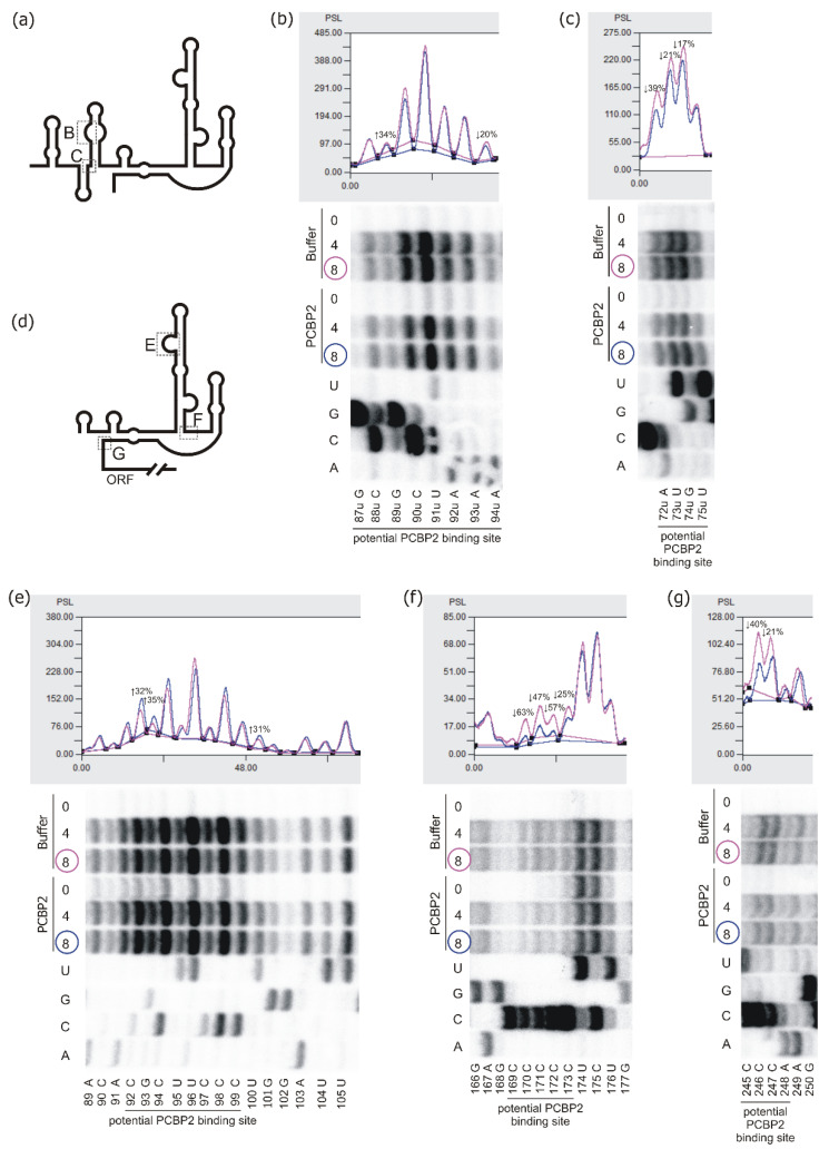
Figure 5.
Structural analysis of the P0-Δ40p53 RNA and p53-554 RNA in the presence of PCBP2 using Pb2+-induced cleavage method. Schematic representation of the secondary structure model of P0-Δ40p53 RNA (a) and p53-554 RNA (d). The analysed regions to which PCBP2 binds are marked with dotted-line boxes B, C, E, F and G. (b,c,e–g) bottom panels: the autoradiograms show the products of Pb2+-induced cleavage reaction identified by reverse transcription method with 5′-end-[32P]-labelled DNA primers. Pb2+-induced cleavage reaction was conducted in the presence of PCBP2 protein or the buffer, at 37 °C for 3 min with Pb2+ ion concentration of 4 mM and 8 mM. Reaction products were separated on 12% polyacrylamide denaturing gels. Sequencing lines are marked A, C, G and U, respectively. Selected nucleotide residues are indicated on the left side of each autoradiogram. Black lines along the gels indicate potential PCBP2 binding sites. Upper panels: normalised reactivities for Pb2+-induced cleavage at Pb2+ ions concentration of 8 mM as a function of nucleotide position.

