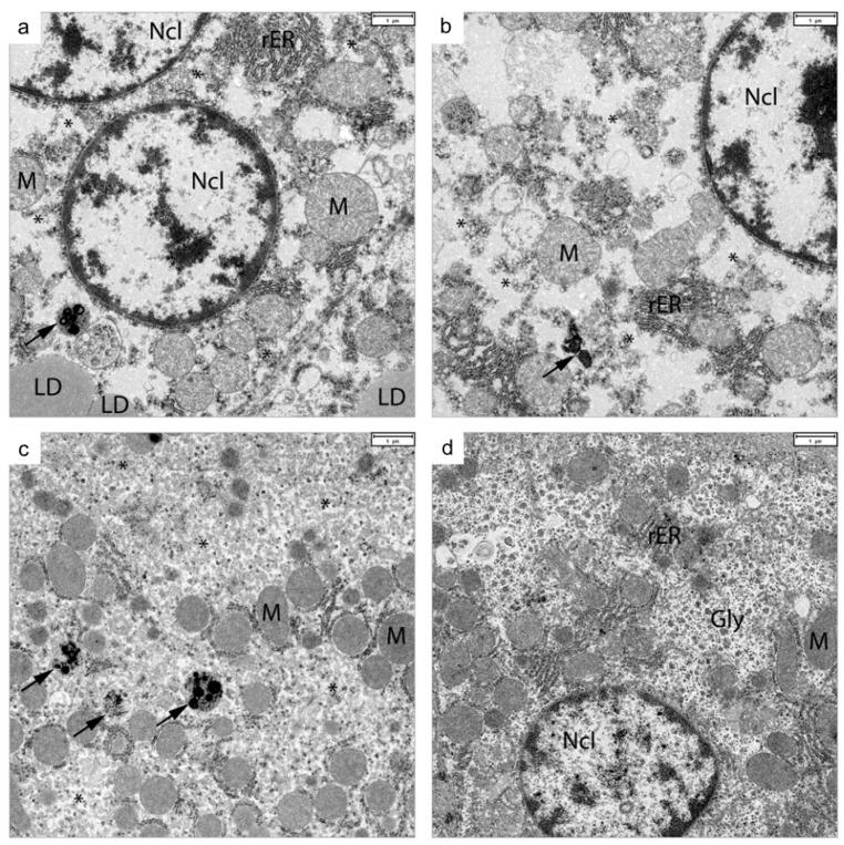
Figure 5.
Transmission electron microscopy of liver samples of p1-FA07 (Hepacivirus F)-infected animals (a–c) compared to a liver sample of a control animal (d), from experiment 4. Hepatocytes of infected bank voles show vesicular endoplasmic reticulum with organoid membranes (*), lipid droplet (LD) accumulation and pleomorphic lysosomal storage vesicles (arrows). Abbreviations: Gly: glycogen rosettes, LD: lipid droplet, M: mitochondria, Ncl: cell nucleus, rER: rough endoplasmic reticulum. Bar in each panel: 1 µm.

