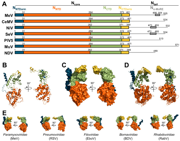
Figure 4.
Organization and structure of the nucleoproteins. (A) Organization and boundaries of the domains of all nucleoproteins whose structure has been solved. Boundaries of Nα-MoRE are only known for MeV [71,73], NiV [74,127], and SeV [128]. (B) Structure of the N protein of MeV in cartoon representations (PDB: 6H5Q). (C) Structure of the N protein of MeV in surface representations (PDB: 6H5Q). (D) Superimposition of aligned structures of MeV (PDB: 6H5Q), CeMV (PDB: 7OI3), NiV (PDB: 7NT5), SeV (PDB: 6M7D), PIV5 (PDB: 4XJN), MuV (PDB: 7EWQ), and NDV (PDB: 6JC3). (E) Surface representations of the structure of the N proteins of MeV (PDB: 6H5Q), respiratory syncytial virus (RSV, PDB: 2WJ8), Ebola virus (EboV, PDB: 5Z9W), Borna disease virus (BDV, PDB: 1N93), and rabies virus (RabV, PDB: 2GTT). All the structures shown in Figure 4 correspond to N proteins observed in their assembled form (i.e., in NCLC).

