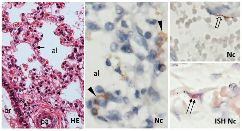
Figure 4.
Lungs of the fetus had normal histological structure of bronchi (br), pulmonary arteries (pa), and alveoli (al); only a slight increase in small lymphocyte infiltrate (arrow) in the interstitium was noted. Small groups or individual alveolar epithelial cells (arrowhead) and a few individual endothelial cells (open arrow) showed immunohistochemical positivity of SARS-CoV-2 nucleocapsid protein (Nc). Scattered endothelial cells were positive for the presence of SARS-CoV-2 RNA by in situ hybridization (ISH; double arrow). HE, 200×; Nc, ISH Nc, 600×.

