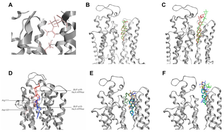
Figure 3.
Cardiotonic steroids-binding site in Na,K-ATPase models. (A)—cardiotonic steroids (CTSs) binding site in α1R-Na,K-ATPase. Hydrogen bonds between Arg111 and Asp122 in α1R-Na,K-ATPase. (B,C)—verification of ouabain and digoxin position in CTSs-binding site of α1S-Na,K-ATPase. Comparison from X-ray 4HYT and 4RET structures with docking models: (B)—ouabain in 4HYT structure, (C)—digoxin in 4RET structure. CTSs from 4HYT or 4RET colored red, CTSs from docking models colored green. (D)—bufalin position in CTSs-binding site of α1S- (blue) and α1R-Na,K-ATPase (red). Models based on the 4HYT structure of Na,K-ATPase. (E,F)—CTSs positions in the CTSs-binding site of α1S-Na,K-ATPase (E) and α1R-Na,K-ATPase (F) for CTS. Bufalin—shown in turquoise, digoxin—green, ouabain—blue, marinobufagenin—orange. Models based on the 4HYT structure of Na,K-ATPase. BUF—bufalin.

