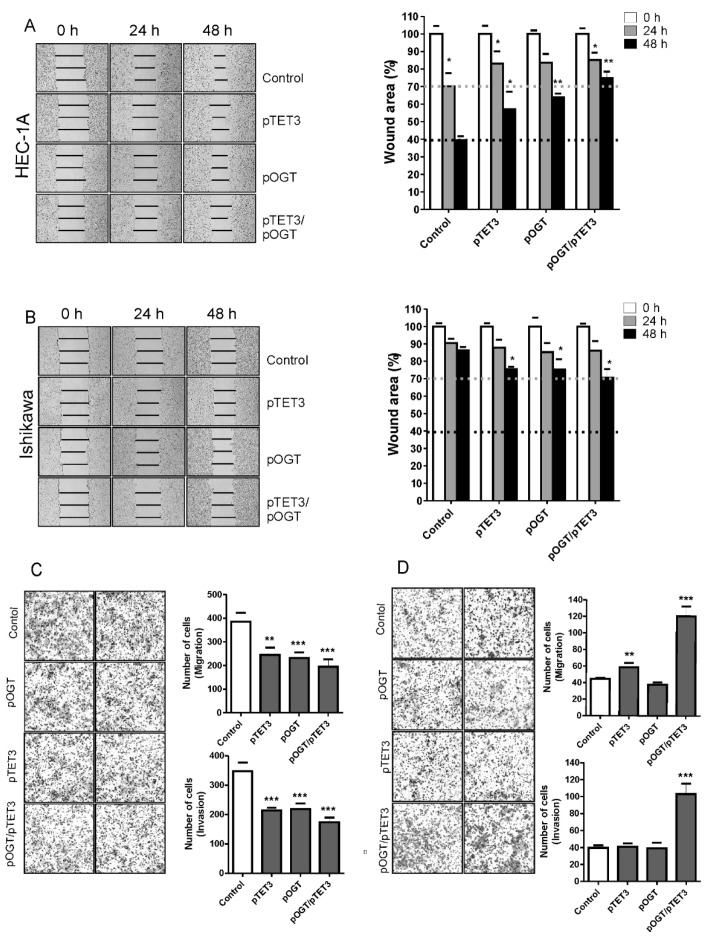
Figure 5.
Effect of OGT and TET3 on migration and invasion of cells. The wound-healing assay was performed to examine the migration rate of HEC-1A (A) and Ishikawa cells (B) transduced with OGT and TET3 plasmid vectors. Photographs were taken at 0, 24 h, and 48 h following the initial scratch. Migration rates were quantified by measuring three different wound areas. Three separate experiments were performed. Cell migration assays using Transwell chambers and invasion assays using Transwell chambers with Matrigel were performed for HEC-1A (C) and Ishikawa cells (D). Representative images of migrating cells stained with Giemsa are displayed (left) for HEC-1A (C) and Ishikawa cells (D). Quantitative data of migration and invasion assay are expressed relative to the migration and invasion abilities of control cells. Plots show average counts from three independent testings * p < 0.01; ** p < 0.001; *** p < 0.0001.

