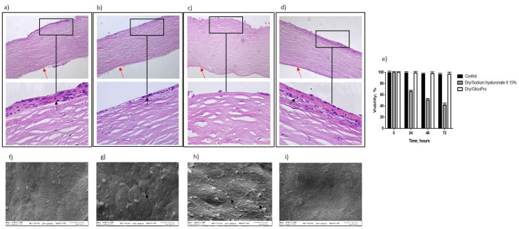
Figure 2.
Histo-morphological analysis. Histo-morphological analysis of (a) control group corneal tissues; (b) dry eye; (c) control dry eye (100 µL sodium hyaluronate 0.15%), and (d) dry eye corneal tissues treated with GlicoPro for 24 h. Magnifications: 10× (upper panels), 60× (lower panel). Red arrows indicated endothelial cell layer; black arrows indicated stratified squamous epithelium. (e) Cell viability assessed by trypan blue staining. Scanning electron microscopy images of human corneal tissue in (f) control; (g) dry eye; (h) control dry eye (100 µL sodium hyaluronate 0.15%); and (i) GlicoPro-treated dry eye conditions. Each condition was analyzed in triplicate. Scale bars = 100 μm.

