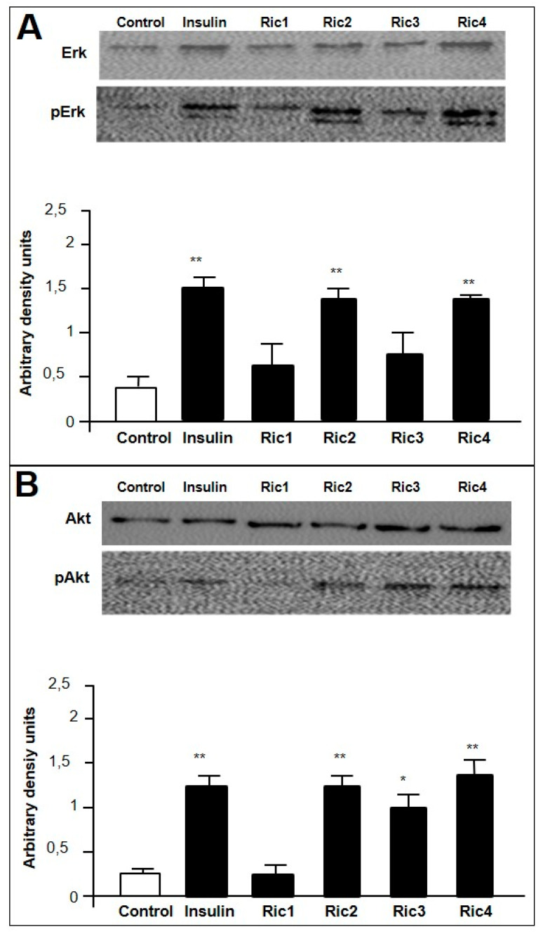
Figure 2.
C2C12 cell signaling assays by Western blotting. C2C12 cells previously differentiated into myotubes were treated in the presence or absence of insulin (100 nM), or the indicated intracellular peptide (Ric1, Ric2, Ric3, or Ric4; 100 µM). Induced phosphorylation of Erk (A) or Akt (B), respectively, pAkt or pErk, were analyzed by Western blotting as described in Section 2.5. Imaging and band intensity measurements were performed using the ImageJ 1.49 software, and quantifications were performed evaluating the relative levels of pErk or pAkt over total Erk or Akt, respectively. The effect of the indicated peptide on the relative phosphorylation levels were expressed using arbitrary density units (A,B, lower panels). Data are representative of three independent experiments that produced similar results. The statistical comparisons were performed using Student’s t-test or analysis of variance (ANOVA), followed by ad-hoc Tukey’s test using GraphPad Prism software * p < 0.05; ** p < 0.001.

